39 brain mri with labels
Brain MRI: How to read MRI brain scan | Kenhub MRI is the most sensitive imaging method when it comes to examining the structure of the brain and spinal cord. It works by exciting the tissue hydrogen protons, which in turn emit electromagnetic signals back to the MRI machine. The MRI machine detects their intensity and translates it into a gray-scale MRI image. › en › e-AnatomyBrain: Atlas of human anatomy with MRI - e-Anatomy - IMAIOS Sep 13, 2021 · MRI Atlas of the Brain. This page presents a comprehensive series of labeled axial, sagittal and coronal images from a normal human brain magnetic resonance imaging exam. This MRI brain cross-sectional anatomy tool serves as a reference atlas to guide radiologists and researchers in the accurate identification of the brain structures.
Head MRI: Purpose, Preparation, and Procedure - Healthline Magnetic resonance imaging (MRI) of the head is a painless, noninvasive test that produces detailed images of your brain and brain stem. An MRI machine creates the images using a magnetic...
Brain mri with labels
MRI-labeling: label human brain MRI image by AAL/BA system Input description: x, y, z : x,y,z value of the mni coordinate. distance (default is T): If the MNI coordinate does not belong to any AAL/BA brain region (e.g. white matter, ventricle), then output the closest AAL/BA brain region name and the their distance (mm). When the MNI coordinate does fall into an AAL brain region, then output distance=0. Brain MRI: What It Is, Purpose, Procedure & Results - Cleveland Clinic A brain MRI (magnetic resonance imaging) scan, also called a head MRI, is a painless procedure that produces very clear images of the structures inside of your head — mainly, your brain. MRI uses a large magnet, radio waves and a computer to produce these detailed images. It doesn't use radiation. Brain lobes - annotated MRI | Radiology Case | Radiopaedia.org Magnetic resonance imaging (MRI) scanning for research: the experiences of healthy volunteers and patients with remitted depressive illness. Victoria Tischler et al., Mental Health Review Journal, 2009. Biomarkers for the diagnosis of Alzheimer's disease, dementia Lewy body, frontotemporal dementia and vascular dementia.
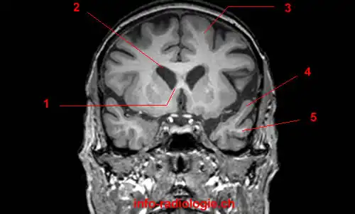
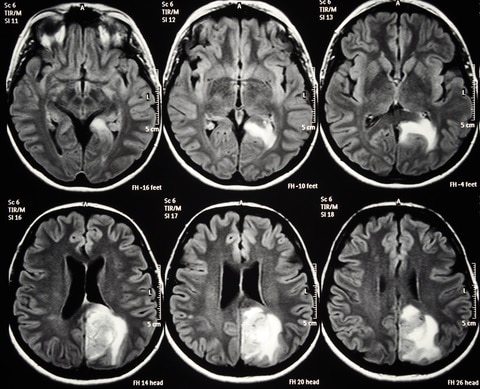
Brain mri with labels. brain anatomy | MRI coronal brain anatomy | free MRI cross sectional ... ELBOW AXIAL. WRIST AXIAL. WRIST CORONAL. KNEE CORONAL. KNEE SAGITTAL. ARTERIES UPPER LEG. ARTERIES LOWER LEG. This MRI brain coronal cross sectional anatomy tool is absolutely free to use. Use the mouse scroll wheel to move the images up and down alternatively use the tiny arrows (>>) on both side of the image to move the images. Brain Tumor MRI Dataset | Kaggle A brain tumor is a collection, or mass, of abnormal cells in your brain. Your skull, which encloses your brain, is very rigid. Any growth inside such a restricted space can cause problems. Brain tumors can be cancerous (malignant) or noncancerous (benign). When benign or malignant tumors grow, they can cause the pressure inside your skull to ... 101 Labeled Brain Images and a Consistent Human Cortical Labeling ... Labeled anatomical subdivisions of the brain enable one to quantify and report brain imaging data within brain regions, which is routinely done for functional, diffusion, and structural magnetic resonance images (f/d/MRI) and positron emission tomography data. Brain MRI segmentation | Kaggle Journal of Neuro-Oncology, 2017. This dataset contains brain MR images together with manual FLAIR abnormality segmentation masks. The images were obtained from The Cancer Imaging Archive (TCIA). They correspond to 110 patients included in The Cancer Genome Atlas (TCGA) lower-grade glioma collection with at least fluid-attenuated inversion ...
en.wikipedia.org › wiki › Spinal_cord_injurySpinal cord injury - Wikipedia CT gives greater detail than X-rays, but exposes the patient to more radiation, and it still does not give images of the spinal cord or ligaments; MRI shows body structures in the greatest detail. Thus it is the standard for anyone who has neurological deficits found in SCI or is thought to have an unstable spinal column injury. A normative spatiotemporal MRI atlas of the fetal brain for automatic ... through manual segmentation the following structures (right and left, when applicable) were labeled on fetal brain mri atlases: hippocampi, amygdala, fornix, cerebellum, brainstem, caudate nuclei, thalami, subthalamic nuclei, lentiform nuclei, corpus callosum, lateral ventricles, developing white matter, cortical plate, and cerebrospinal fluid … yeson30.org › aboutAbout Our Coalition - Clean Air California About Our Coalition. Prop 30 is supported by a coalition including CalFire Firefighters, the American Lung Association, environmental organizations, electrical workers and businesses that want to improve California’s air quality by fighting and preventing wildfires and reducing air pollution from vehicles. Labeled imaging anatomy cases | Radiology Reference Article ... This article lists a series of labeled imaging anatomy cases by body region and modality. Brain CT head: non-contrast axial CT head: non-contrast coronal CT head: non-contrast sagittal CT head: angiogram axial CT head: angiogram coronal CT...
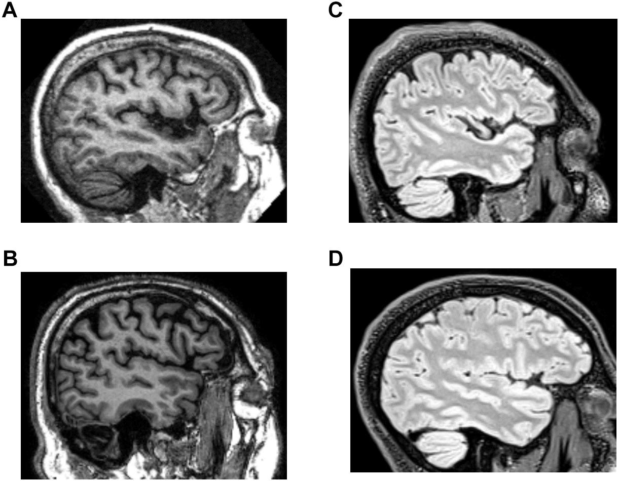
Anatomy, medical imaging and e-learning for healthcare IMAIOS and selected third parties, use cookies or similar technologies, in particular for audience measurement. Cookies allow us to analyze and store information such as the characteristics of your device as well as certain personal data (e.g., IP addresses, navigation, usage or geolocation data, unique identifiers). › Lower-Limb › Ankle-Foot-MRIAnatomy of the foot and ankle - MRI - e-Anatomy - IMAIOS Aug 26, 2022 · Cross-sectional anatomy: MRI of the ankle and feet A magnetic resonance imaging (MRI) was performed on a normal subject; with spin-echo T1 weighted images and spin-echo fat-saturated proton density weighted images (3 usual planes used for osteo-articular imaging: axial, coronal, and sagittal). MRI head sagittal T1 - labeling questions | Radiology Case ... The labeled structures are (excluding the correct side): temporal horn of lateral ventricle primary fissure of cerebellum choroid plexus trigone (atrium) of lateral ventricle horizontal fissure of cerebellum occipital horn of lateral ventricle intraorbital segment of optic nerve diploic space of parietal bone body of caudate nucleus maxillary sinus Brainstem Anatomy - W-Radiology The brainstem refers to the middle part of the brain (1). It consists of the medulla, pons, and midbrain. The brainstem helps relay sensory information, such as pain, eye and mouth movement, involuntary muscle movements, consciousness, respirations, hunger, and cardiac function (2). These functions are possible because of the brainstem's ...
Brain Imaging in Multiple Sclerosis: Practice Essentials ...
Magnetic Resonance Imaging (MRI) of the Spine and Brain Magnetic resonance imaging (MRI) is a diagnostic procedure that uses a combination of a large magnet, radiofrequencies, and a computer to produce detailed images of organs and structures within the body. Unlike X-rays or computed tomography (CT scans), MRI does not use ionizing radiation. Some MRI machines look like narrow tunnels, while others ...
Frontiers | DeepNavNet: Automated Landmark Localization for ...
Brain MRI Images for Brain Tumor Detection | Kaggle Brain MRI Images for Brain Tumor Detection. Brain MRI Images for Brain Tumor Detection. Data. Code (250) Discussion (8) About Dataset. No description available. Health Biology Classification Computer Vision Deep Learning. Edit Tags. close. search. Apply up to 5 tags to help Kaggle users find your dataset.
The Radiology Assistant : Anatomy
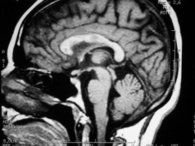
Atlas of BRAIN MRI - W-Radiology Brain magnetic resonance imaging (MRI) is a common medical imaging method that allows clinicians to examine the brain's anatomy (1). It uses a magnetic field and radio waves to produce detailed images of the brain and the brainstem to detect various conditions (2).

Label Each Part of the Brain Scan | MS in African Americans ...
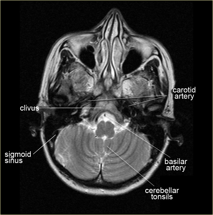
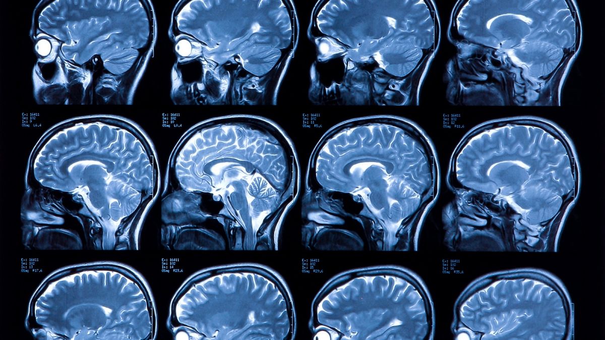
› en › e-AnatomyCross-sectional anatomy of the brain - e-Anatomy - IMAIOS Apr 15, 2022 · Axial MRI Atlas of the Brain. Free online atlas with a comprehensive series of T1, contrast-enhanced T1, T2, T2*, FLAIR, Diffusion -weighted axial images from a normal humain brain. Scroll through the images with detailed labeling using our interactive interface. Perfect for clinicians, radiologists and residents reading brain MRI studies.
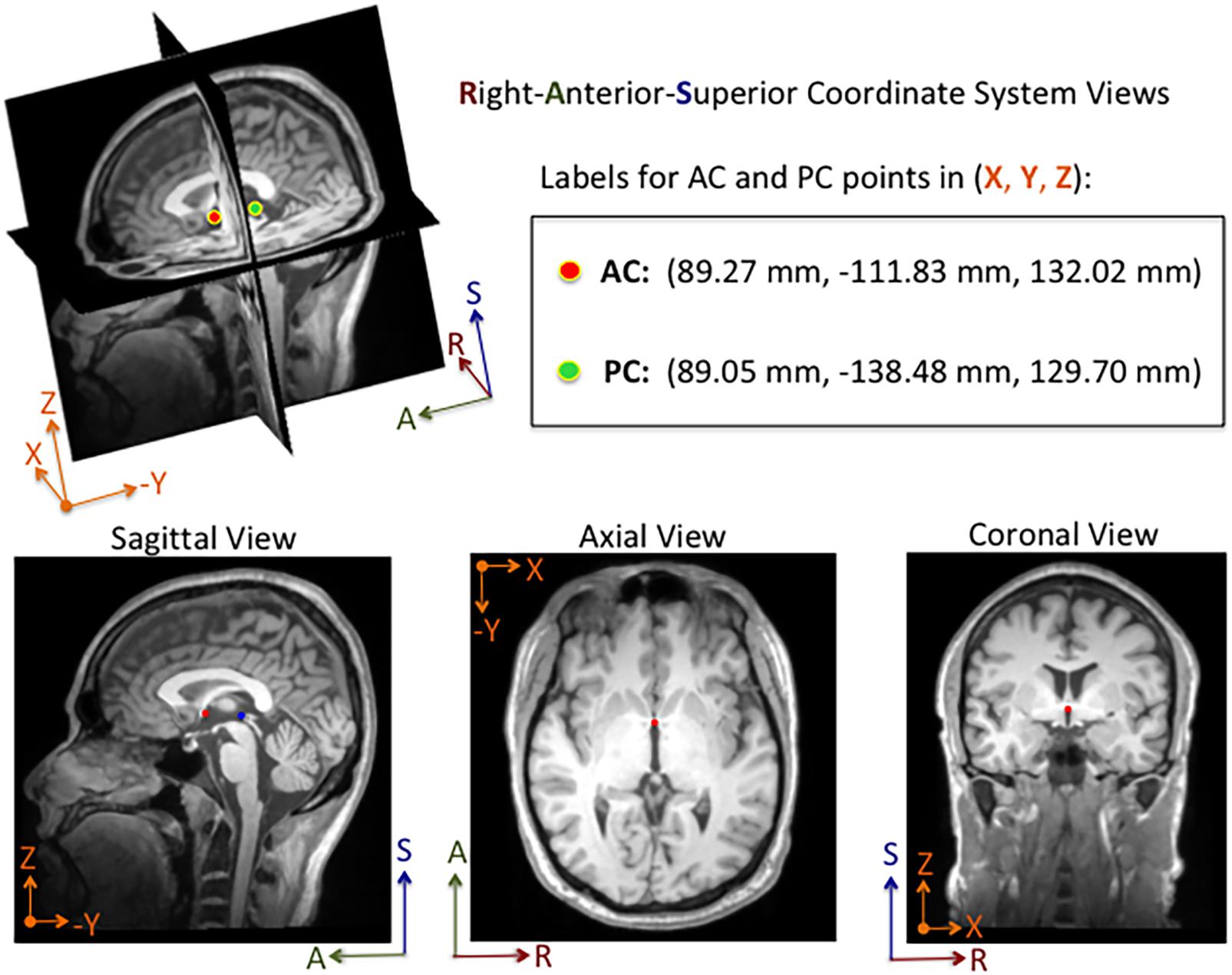
Orientation and Voxel-Order Terminology: RAS, LAS, LPI, RPI ...
› articles › s41598/021/90428-8Brain tumor segmentation based on deep learning and an ... May 25, 2021 · Brain tumor localization and segmentation from magnetic resonance imaging (MRI) are hard and important tasks for several applications in the field of medical analysis. As each brain imaging ...

Brain Anatomy MRI- Neuroradiology
Labeled MRI Brain Scans - Neuromorphometrics We can also label scans that you provide and we are very interested in labeling white matter anatomy as seen in diffusion-weighted MRI scans. If you want an aggregate version of our data, we can provide it as a probabilistic atlas. The cost to label a single scan is $2449 (USD).

volBrain: Automated MRI Brain volumetry system
MRI Brain Animated Quiz - University of Minnesota Canine Brain MRI Anatomy Quiz Sequentially click/tap: first the dot associated with a term; then, its corresponding target dot on the MRI image. If a line connection appears, your choice was correct! White Matter Cerebral Cortex Olfactory Bulb Longitudinal Fissure Levels ROSTRAL

Brain lobes - annotated MRI | Radiology Case | Radiopaedia.org
101 labeled brain images and a consistent human cortical ... - PubMed We introduce the Mindboggle-101 dataset, the largest and most complete set of free, publicly accessible, manually labeled human brain images. To manually label the macroscopic anatomy in magnetic resonance images of 101 healthy participants, we created a new cortical labeling protocol that relies on robust anatomical landmarks and minimal manual edits after initialization with automated labels.

Head and spine anatomy - Radiology Cafe
MRI brain (summary) | Radiology Reference Article - Radiopaedia MRI brain is a specialist investigation that is used for the assessment of a number of neurological conditions. It is the main method to investigate conditions such as multiple sclerosis and headaches, and used to characterize strokes and space-occupying lesions. Reference article

NeuroRad for iPad is a great app for medical professionals to ...
Automated MRI image labelling processes 100,000 brain exams in under 30 ... Now, more than 100,00 MRI examinations can be labeled in less than half an hour. Published in European Radiology, this is the first study allowing researchers to label complex MRI image datasets ...

brain anatomy | MRI coronal brain anatomy | free MRI cross ...
› health-news › is-it-safe-toIs It Safe to Undergo Multiple MRI Exams? - Healthline Sep 27, 2018 · The findings, at the very least, are a cause for concern. That’s what Dr. Emanuel Kanal says about the Food and Drug Administration’s safety announcement last week on the risk of brain ...
Design and fabrication of a realistic anthropomorphic ...
e-Anatomy: radiologic anatomy atlas of the human body - e-Anatomy - IMAIOS IMAIOS and selected third parties, use cookies or similar technologies, in particular for audience measurement. Cookies allow us to analyze and store information such as the characteristics of your device as well as certain personal data (e.g., IP addresses, navigation, usage or geolocation data, unique identifiers).

How To Read A Brain MRI Radiology Report - Part II | Blog ...
UCLA Brain Mapping Center - ICBM Template The ICBM (International Consortium for Brain Mapping) high-resolution single subject template is aligned with the individual subject T1 whole brain MRI provided. An AIR nonlinear warp, a 5th order polynomial, is used for this fit. The demarcated labels on the template are then resampled through the warp transform to the subject MRI.
Magnetic resonance image (MRI) of a side view of the brain ...
What Does a Brain MRI Show? • San Diego Health What does a brain MRI show? The answer is, unfortunately, not very. MRI scans (magnetic resonance imaging) have been around for decades, and the technology has been steadily improving. Today, a brain MRI test can identify whether or not a person has a stroke, or if the person has suffered a traumatic brain injury, or if the person is suffering ...

MRI Scans Show The Horrific Effect Cocaine Abuse Can Have On ...
Frontiers | 101 Labeled Brain Images and a Consistent Human Cortical ... Labeled anatomical subdivisions of the brain enable one to quantify and report brain imaging data within brain regions, which is routinely done for functional, diffusion, and structural magnetic resonance images (f/d/MRI) and positron emission tomography data.

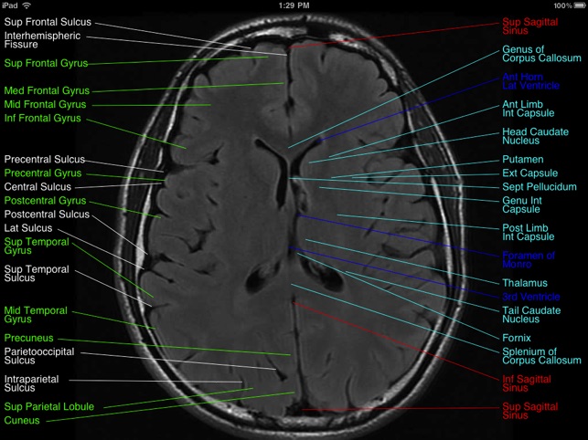
MRI anatomy | free MRI axial brain anatomy
MRI anatomy | free MRI axial brain anatomy - Mrimaster.com This MRI brain cross sectional anatomy tool is absolutely free to use. Use the mouse scroll wheel to move the images up and down alternatively use the tiny arrows (>>) on both side of the image to move the images.

Region Of Interest Based Image Classification: A Study in MRI ...
What to Expect in an MRI for the Head and the Brain A head MRI is a noninvasive imaging test that creates detailed pictures of your brain and surrounding tissues. An MRI allows your doctor to see inside your brain to check for diseases or injuries without having to do surgery. Your doctor can use the images to make a diagnosis and recommend the best treatment for your condition.

Brain imaging in MS,
3 steps to optimize your MRI protocol
Brain lobes - annotated MRI | Radiology Case | Radiopaedia.org Neuro- MRI by Dr Bálint Botz Head MRI by Anthony Pennuto; Annotated CT/MR Teaching by Matt Wong Brain Anatomy MRI by R. Furman Borst MD; fälle für Anatomie by Eva Fischer NRAD by Johann Jende; Частки ГМ 2021 by Василь; Annotated Anatomy by Marc Hidalgo; 6_NEUROLOGIC IMAGING - Weissleder by Felicia Wright; MRI BRAIN by Dr ...

Brain ventricle parcellation using a deep neural network ...
Brain lobes - annotated MRI | Radiology Case | Radiopaedia.org Magnetic resonance imaging (MRI) scanning for research: the experiences of healthy volunteers and patients with remitted depressive illness. Victoria Tischler et al., Mental Health Review Journal, 2009. Biomarkers for the diagnosis of Alzheimer's disease, dementia Lewy body, frontotemporal dementia and vascular dementia.

volBrain: Automated MRI Brain volumetry system
Brain MRI: What It Is, Purpose, Procedure & Results - Cleveland Clinic A brain MRI (magnetic resonance imaging) scan, also called a head MRI, is a painless procedure that produces very clear images of the structures inside of your head — mainly, your brain. MRI uses a large magnet, radio waves and a computer to produce these detailed images. It doesn't use radiation.

Labelled MRI of Normal Brain - Stock Image - C017/4418 ...
MRI-labeling: label human brain MRI image by AAL/BA system Input description: x, y, z : x,y,z value of the mni coordinate. distance (default is T): If the MNI coordinate does not belong to any AAL/BA brain region (e.g. white matter, ventricle), then output the closest AAL/BA brain region name and the their distance (mm). When the MNI coordinate does fall into an AAL brain region, then output distance=0.

Brain Tumor Detection and Localization - Analytics Vidhya

Brain: Atlas of human anatomy with MRI - e-Anatomy
Brain MRI Atlas on the App Store

Automated segmentation of the hypothalamus and associated ...

Arterial Spin-Labeling Improves Detection of Intracranial ...

Labelled MRI of Normal Brain - Stock Image - C017/4421 ...

Tips and traps in brain MRI: Applications to vascular ...
Atlas of BRAIN MRI - W-Radiology

Cross-sectional anatomy of the brain - e-Anatomy
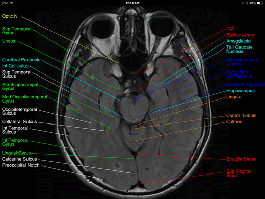
Review of “Brain MRI Atlas” App for the iPad | SpringerLink
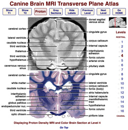
Veterinary Planar Anatomy Courseware
Frontiers | Systematic Differences Between Perceptually ...

Early postmortem brain MRI findings in COVID-19 non-survivors ...

MRI anatomy | free MRI axial brain anatomy

Cross-sectional anatomy of the brain - e-Anatomy
Atlas of BRAIN MRI - W-Radiology
Potentially life-saving study could cut labelling times for ...
Brain Lesion Detection in MRI Images with Graph-cut Algorithms
Post a Comment for "39 brain mri with labels"