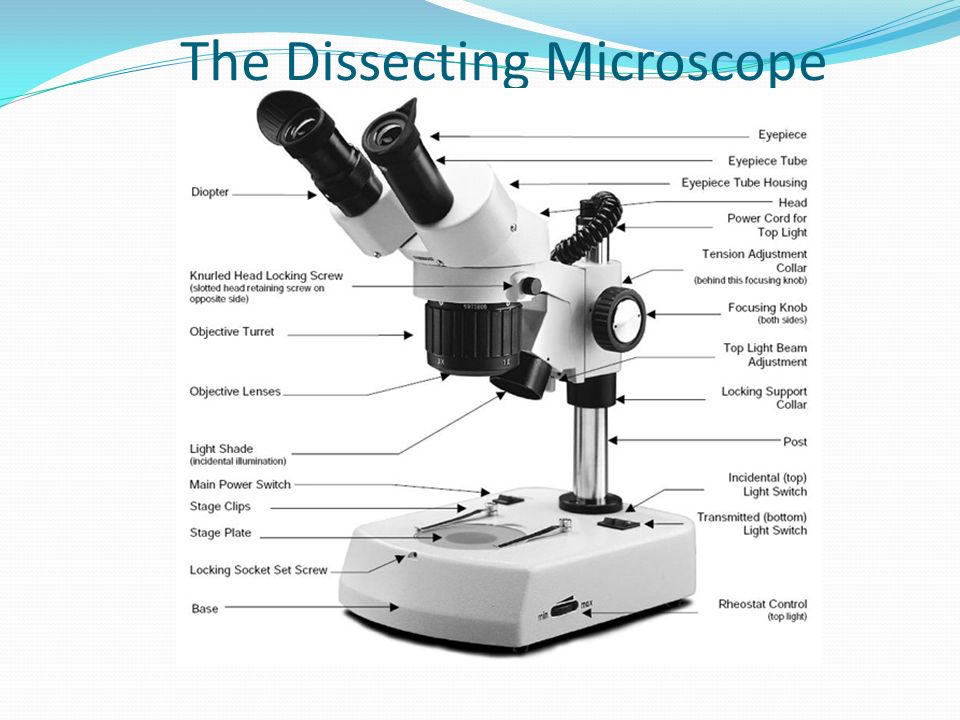
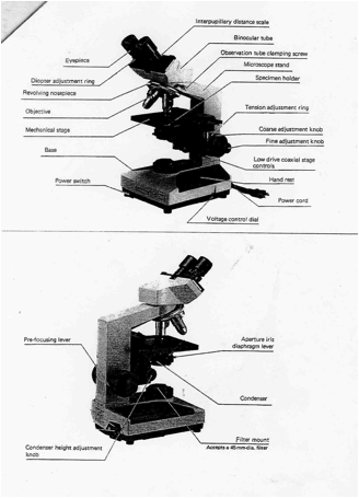
44 dissecting microscope diagram with labels
Microscope Parts and Functions First, the purpose of a microscope is to magnify a small object or to magnify the fine details of a larger object in order to examine minute specimens that cannot be seen by the naked eye. Here are the important compound microscope parts... Eyepiece: The lens the viewer looks through to see the specimen. A Study of the Microscope and its Functions With a Labeled Diagram ... A Study of the Microscope and its Functions With a Labeled Diagram To better understand the structure and function of a microscope, we need to take a look at the labeled microscope diagrams of the compound and electron microscope. These diagrams clearly explain the functioning of the microscopes along with their respective parts.
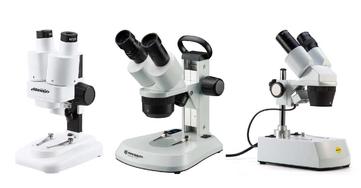
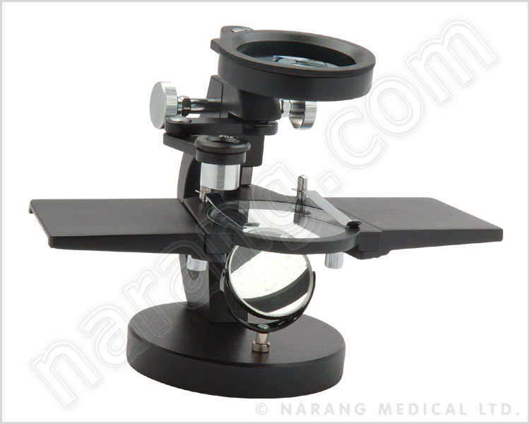
Parts of the Dissecting Microscope - Synonym Dissecting microscopes are used for viewing live specimens or three-dimensional objects too large or thick to be accommodated by compound microscopes. Specimens can be physically manipulated under magnification, since they do not have to be mounted onto a slide for observation under a dissecting microscope. These ...
Dissecting microscope diagram with labels
Parts of a microscope with functions and labeled diagram - Microbe Notes Figure: Diagram of parts of a microscope There are three structural parts of the microscope i.e. head, base, and arm. Head - This is also known as the body. It carries the optical parts in the upper part of the microscope. Base - It acts as microscopes support. It also carries microscopic illuminators. Hot and Cold Packs: A Thermochemistry Activity | Carolina.com Diagram your hot or cold pack. Include labels to indicate sizes and quantities of materials used. List all materials and quantities needed to create your thermal pack. Explain the steps that you will follow to build your thermal pack. Describe the safety precautions you will use when creating and testing the thermal pack. microbiomejournal.biomedcentral.com › articles › 10Gut-derived metabolites influence neurodevelopmental gene ... Aug 23, 2022 · Live larvae were anesthetized in 0.04% tricaine, embedded in 2% methyl cellulose, and imaged with dissecting microscope (V8 Zeiss) mounted with a MicroPublisher 5.0 camera and imaged using Q-Capture software (v 3.1.3.10). Fluorescent images were captured using a Leica CLSM SP5 confocal microscope using LAS AF imaging software v2.7.7.
Dissecting microscope diagram with labels. rsscience.com › stereo-microscopeParts of Stereo Microscope (Dissecting microscope) – labeled ... Stereo microscopes (also called Dissecting microscope) are branched out from other light microscopes for the application of viewing "3D" objects. These include substantial specimens, such as insects, feathers, leaves, rocks, sand grains, gems, coins, and stamps, etc. Functionally, a stereo microscope is like a powerful magnifying glass. Single-cell transcriptome profiling reveals neutrophil ... - Nature Jul 27, 2020 · Specifically, cells would receive corresponding labels with the highest similarity scores, whereas cells with the highest similarity score lower than 0.5 were defined as unassigned. MIT - Massachusetts Institute of Technology a aa aaa aaaa aaacn aaah aaai aaas aab aabb aac aacc aace aachen aacom aacs aacsb aad aadvantage aae aaf aafp aag aah aai aaj aal aalborg aalib aaliyah aall aalto aam ... Label the microscope — Science Learning Hub Use this with the Microscope parts activity to help students identify and label the main parts of a microscope and then describe their functions. Drag and drop the text labels onto the microscope diagram. If you want to redo an answer, click on the box and the answer will go back to the top so you can move it to another box.
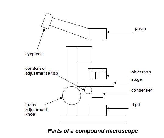
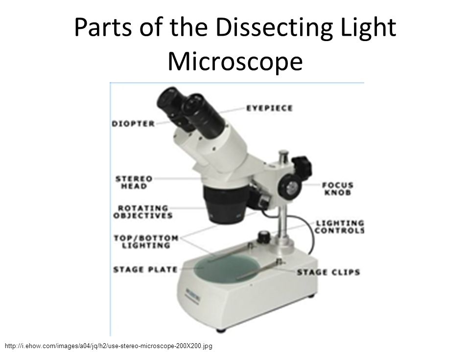
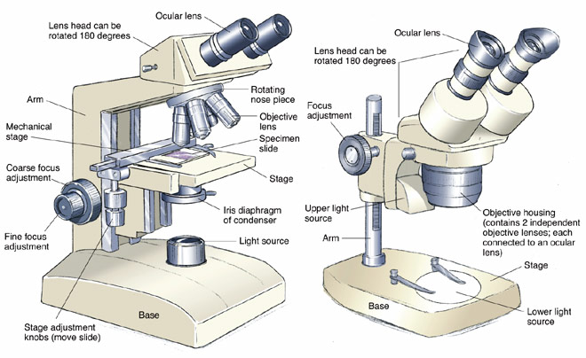
› articles › s41467/022/28497-0The spatial transcriptomic landscape of the healing mouse ... Feb 11, 2022 · The experimental slide with colonic tissue from d0 and d14 was fixed and stained with hematoxylin and eosin (HE) and imaged using a Leica DM5500 B microscope (Leica Microsystems) at 5X magnification. Compound Microscope Parts - Labeled Diagram and their Functions The term "compound" refers to the microscope having more than one lens. Basically, compound microscopes generate magnified images through an aligned pair of the objective lens and the ocular lens. In contrast, "simple microscopes" have only one convex lens and function more like glass magnifiers. [In this figure] Two "antique ... K To 12 Science Grade 7 Learners Material - Module Read and do the activities in the section on How to Use The Light Microscope before performing Activity 2. Activity 2 Investigating plant cells Objectives In this activity, you should be able to: 1. prepare a wet mount; 2. describe a plant cell observed under the … › dissecting-stereoDissecting Stereo Microscope Parts and Functions Dissecting Stereo Microscope Parts and Functions Overview. Also known as a stereoscopic microscope, a dissecting microscope is a type of optical microscope commonly used for studying three-dimensional objects (3-D objects) as well as for dissecting biological specimen (e.g. insects and plant parts etc) at low magnification, between 2 and 100x depending on the microscope.
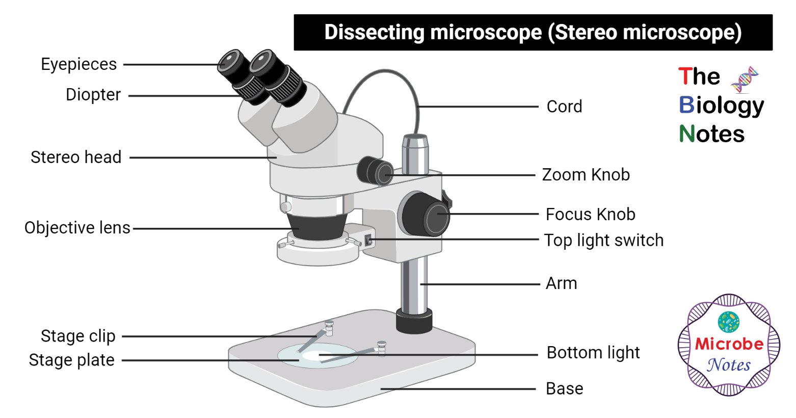
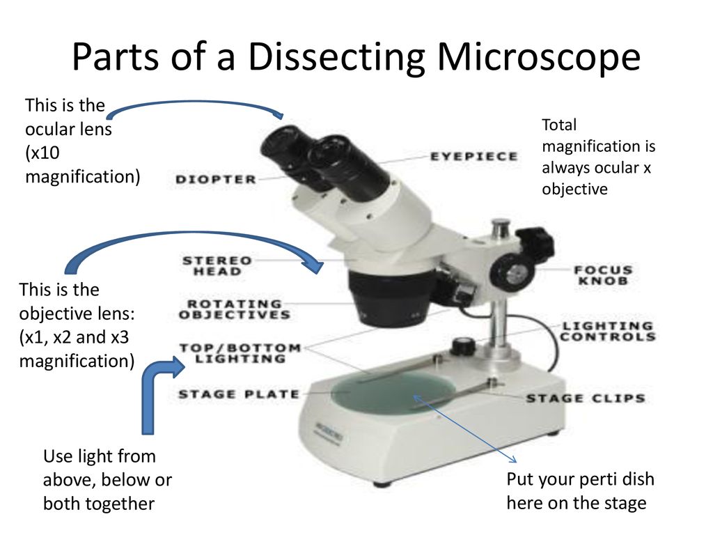
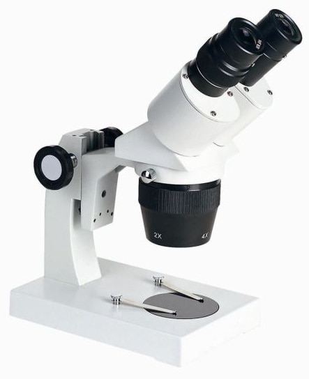
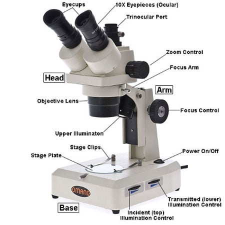
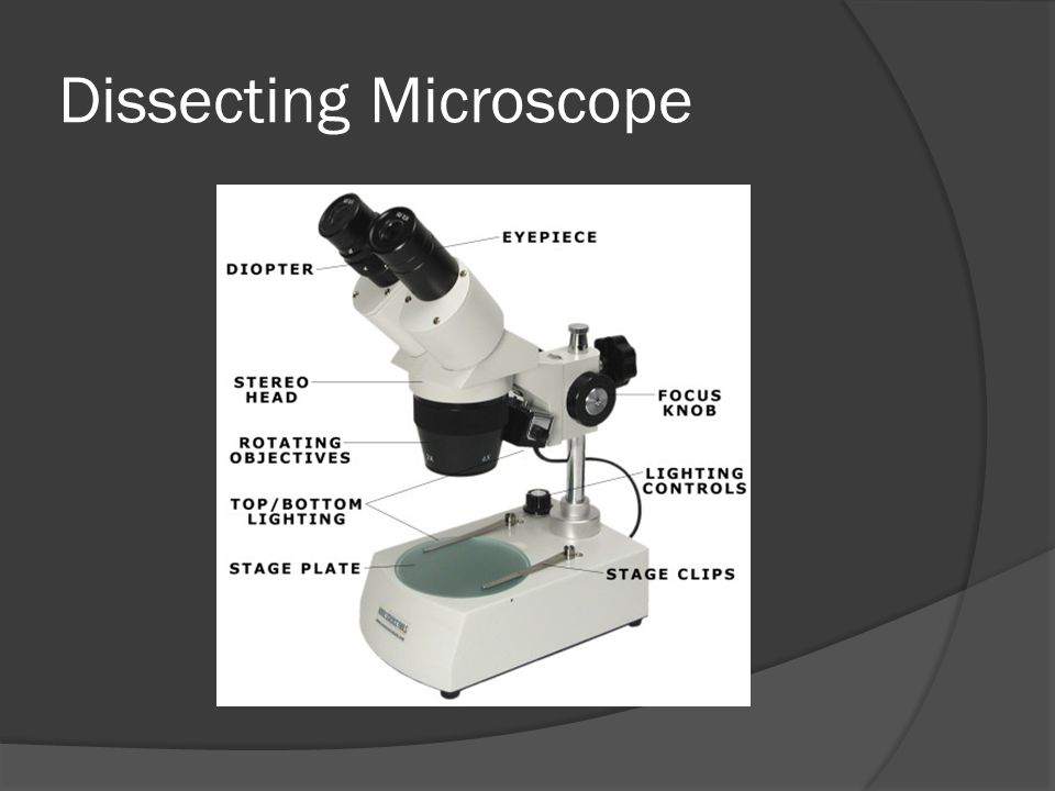
› doi › fullProbing cell identity hierarchies by fate titration and ... Sep 15, 2022 · Using a dissection microscope (Leica), heads, limbs, vertebral columns, and internal organs were removed to make certain no multipotent cells were present in cultures. After dissection, 2–3 embryos were pooled, and tissue was dissociated in 0.15% Trypsin (Gibco) for 10–15 min to obtain single-cell suspensions. Dissecting microscope (Stereoscopic or Stereo microscope) This microscope is a dual-powered dissecting microscope of 10x-30x with an ability to rotate 360° making it ideal for viewing and focussing better to view samples. By rotating the lenses, users can change the magnification of image. Dissecting Microscope Parts And Functions. All You Need To Know The dissecting microscope is also referred to as a stereoscopic microscope and is ordinarily used to study three-dimensional objects. And also as the name suggests for dissecting and analysing biological specimens under low magnification between two and two hundred and fifty times. Parts of Stereo Microscope (Dissecting microscope) – labeled diagram ... Labeled part diagram of a stereo microscope ... (based on color bands and their respective labels), the objectives of a dissecting microscope are located in a cylindrical cone and, therefore, are not directly seen. For the stereo microscope that comes with multiple objective lens sets (fixed power style), the cylindrical cone can be turned to ...
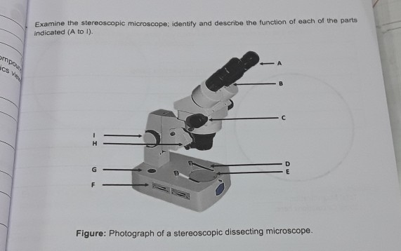
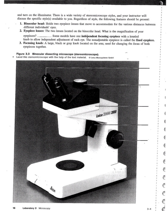
Solved Examine the stereoscopic microscope; identify and ...
Conserved cell types with divergent features in human versus … Aug 21, 2019 · After staining, sections were visualized on a fluorescence dissecting microscope (Leica) and cortical layers were individually microdissected using a needle blade micro-knife (Fine Science Tools ...
Dissecting microscope (Stereo or stereoscopic microscope ...
› createJoin LiveJournal Password requirements: 6 to 30 characters long; ASCII characters only (characters found on a standard US keyboard); must contain at least 4 different symbols;
Microscopes. (a) Binocular dissecting microscope. (b ...
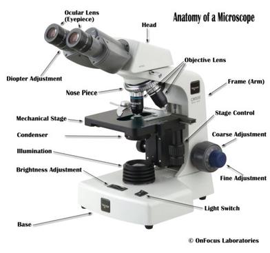
Microscope, Microscope Parts, Labeled Diagram, and Functions Stage with Stage Clips: The stage of a microscope is a flat platform where you place your subject slides. Stage clips hold the slides in place. The mechanical stage of your microscope will help you to move the slide around by turning two knobs. One knobs moves it left and right, the other knobs moves it up and down.
Dissecting Stereo Microscope Parts and Functions
Microscope Label Diagram - 34 label diagram of microscope labels ... Microscope Label Diagram - 18 images - unbiology6, biology 521 resources, label a microscope teaching resources, quia protist vocabulary, Menu ≡ ╳ Home

Basic Microscopy - An Important Skill for Plant Pathologists
Simple Microscope - Diagram (Parts labelled), Principle, Formula and Uses Simple microscope is a magnification apparatus that uses a combination of double convex lens to form an enlarged, erect image of a specimen. The working principle of a simple microscope is that when a lens is held close to the eye, a virtual, magnified and erect image of a specimen is formed at the least possible distance from which a human eye ...
Microscope Work | The British Lichen Society
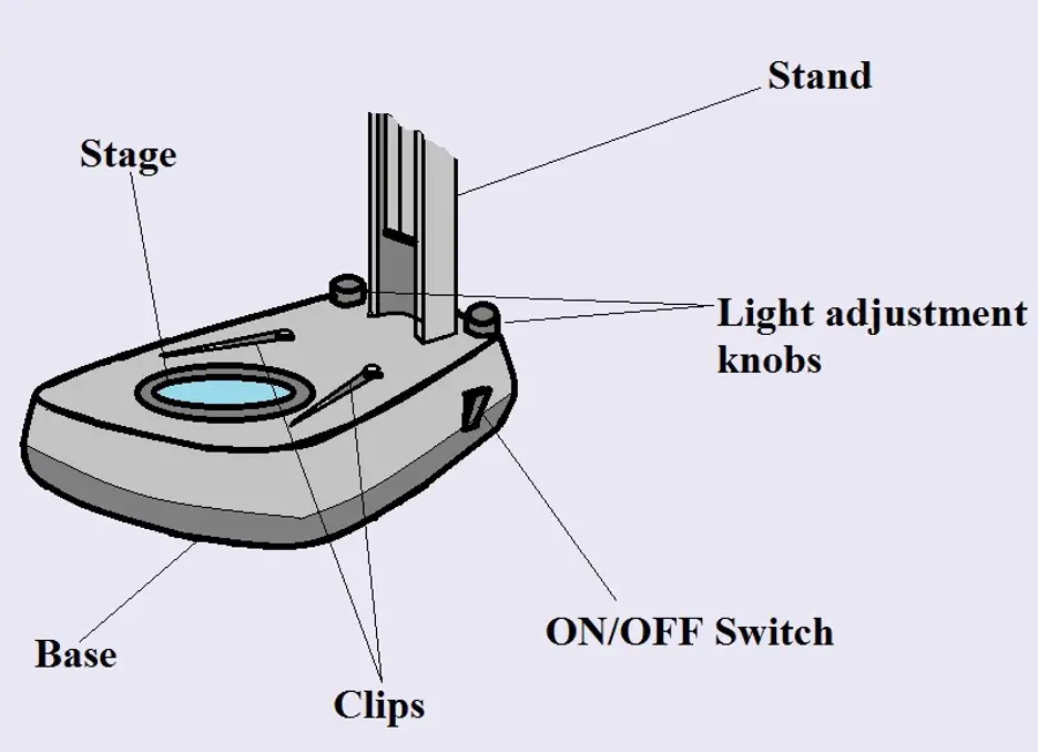
Parts of Dissecting Microscope | Botany - Biology Discussion Dissecting microscope is used to dissect small organisms or organs, e.g., embryo dissection. Its special utility is to observe such materials where high magnification is not needed. Design of Compound Microscope (With Diagram) | Biology Labelled Diagram of Compound Microscope
Parts of Stereo Microscope (Dissecting microscope) – labeled ...
Compound Light/Dissecting Microscope Diagram | Quizlet Used to examine material mounted on microscope slides (usually thinly sectioned & stained) Provides total magnification of 40x-1000x No space for dissection Rules TRANSPORT Arm & base USE Always start at 4x, Coarse focus, Fine focus Then change objectives & use fine focus as needed Coarse focus ONLY with 4x! CLEANING Objectives/Oculars

Stereo Zoom Microscope - Types Of Microscopes | Stereo ...
Microscope labeled diagram - SlideShare Microscope labeled diagram 1. The Microscope Image courtesy of: Microscopehelp.com Basic rules to using the microscope 1. You should always carry a microscope with two hands, one on the arm and the other under the base. 2. You should always start on the lowest power objective lens and should always leave the microscope on the low power lens ...
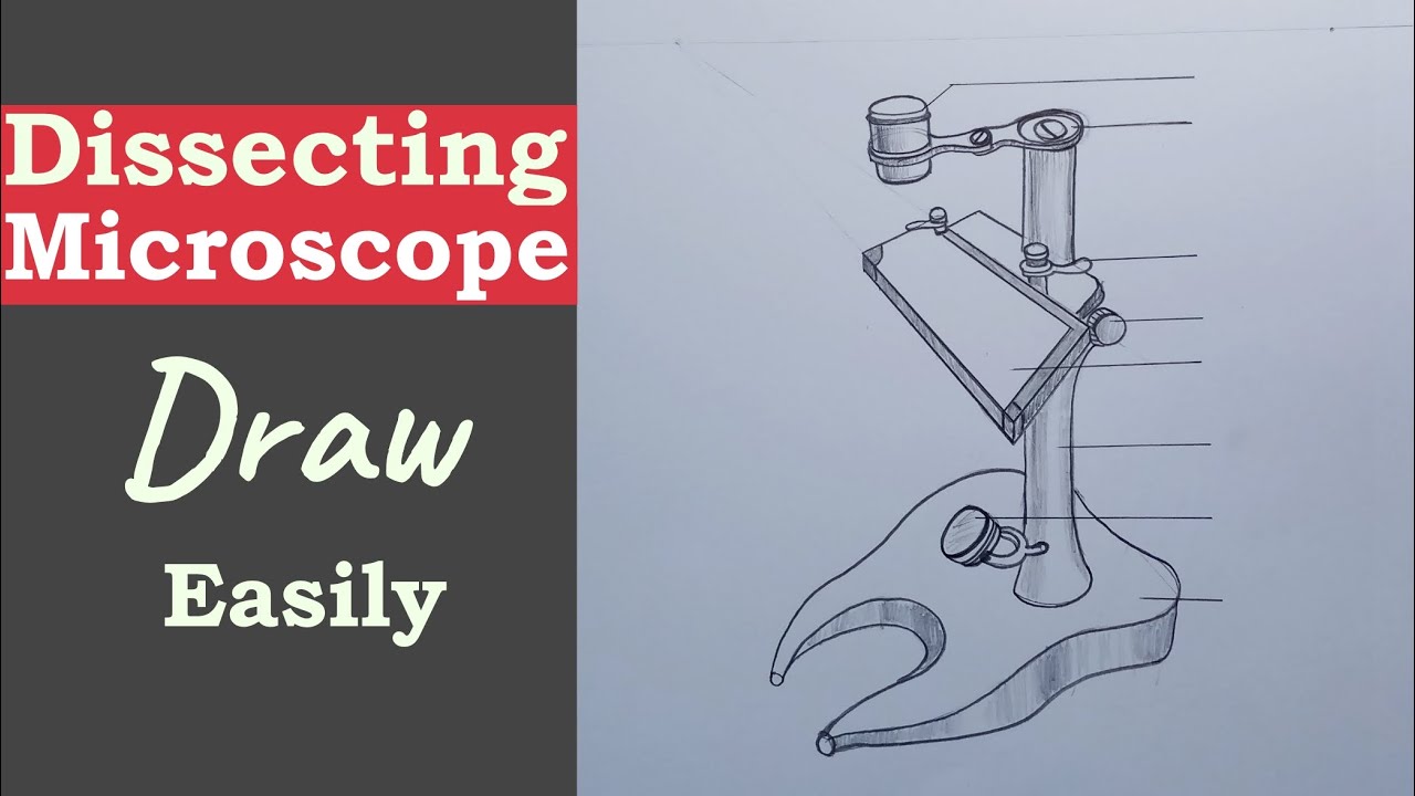
How to Draw a Dissecting Microscope || Dissecting Microscope Drawing || microscope drawing
› articles › s41556/022/00961-5Single-cell analysis of endometriosis reveals a coordinated ... Jul 21, 2022 · Using single-cell analysis, Tan et al. map the cellular and spatial hierarchy and heterogeneity of eutopic endometrium and characterize ectopic peritoneal and ovarian endometriosis lesions from ...

How To Draw A Microscope, Step by Step, Drawing Guide, by ...
Dissecting microscope parts and functions pdf - Canada Guid Step-by ... Stereo Microscope (dissecting microscope): These microscopes magnify up to about maximum 100x and supply a 3-dimensional view of the specimen. They are useful for observing opaque objects. They are useful for observing opaque objects. Identify the parts of the compound microscope and explain the function of each part. 5.
Microscopy and Cytology - ppt download
Compound Microscope Parts, Functions, and Labeled Diagram Compound Microscope Definitions for Labels. Eyepiece (ocular lens) with or without Pointer: The part that is looked through at the top of the compound microscope. Eyepieces typically have a magnification between 5x & 30x. Monocular or Binocular Head: Structural support that holds & connects the eyepieces to the objective lenses.

Types of Microscopes: Definition, Working Principle, Diagram ...
Dissecting microscope (Stereo or stereoscopic microscope)- Definition ... Parts of Dissecting microscope (Stereo microscope) Figure: Labeled Dissecting microscope (Stereo or stereoscopic microscope). Image created using biorender.com LED illuminators- For some of the dissecting Microscopes, they have an inbuilt LED illuminator as a source of light.
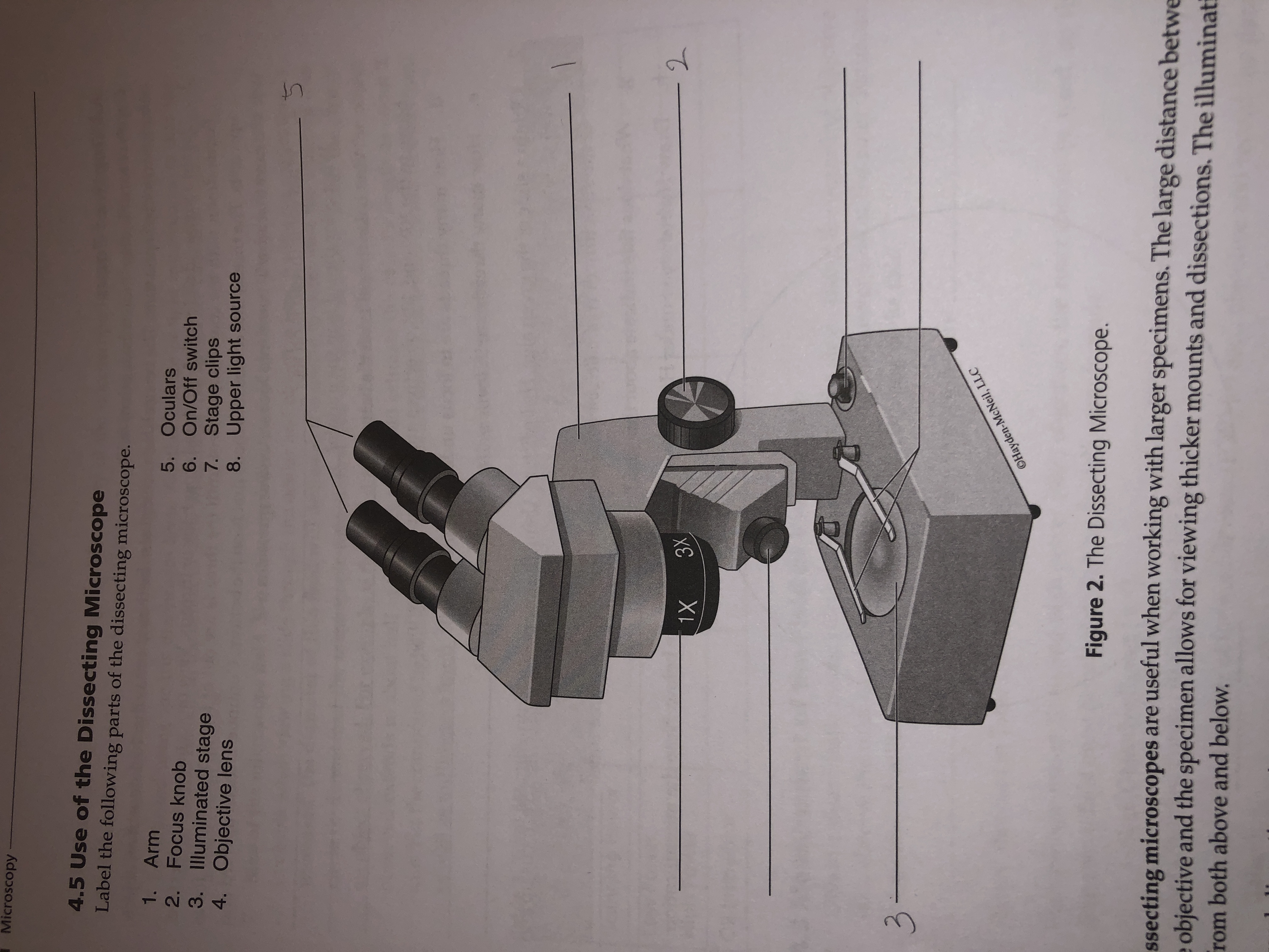
Answered: Microscopy 4.5 Use of the Dissecting… | bartleby
Microscope Types (with labeled diagrams) and Functions These microscopes work on the principle called contrast-enhancing technique that is utilized to produce high-contrast images to view them with more accuracy and clarity. Phase-contrast microscope labeled diagram Phase-contrast microscope functions: Its applications areas include In cases where the specimen is colorless and is very tiny

A Study of the Microscope and its Functions With a Labeled ...
microbiomejournal.biomedcentral.com › articles › 10Gut-derived metabolites influence neurodevelopmental gene ... Aug 23, 2022 · Live larvae were anesthetized in 0.04% tricaine, embedded in 2% methyl cellulose, and imaged with dissecting microscope (V8 Zeiss) mounted with a MicroPublisher 5.0 camera and imaged using Q-Capture software (v 3.1.3.10). Fluorescent images were captured using a Leica CLSM SP5 confocal microscope using LAS AF imaging software v2.7.7.

Microscope World Blog: Dissecting Microscopes
Hot and Cold Packs: A Thermochemistry Activity | Carolina.com Diagram your hot or cold pack. Include labels to indicate sizes and quantities of materials used. List all materials and quantities needed to create your thermal pack. Explain the steps that you will follow to build your thermal pack. Describe the safety precautions you will use when creating and testing the thermal pack.
Dissecting Microscopes | Senior Dissecting Microscopes ...
Parts of a microscope with functions and labeled diagram - Microbe Notes Figure: Diagram of parts of a microscope There are three structural parts of the microscope i.e. head, base, and arm. Head - This is also known as the body. It carries the optical parts in the upper part of the microscope. Base - It acts as microscopes support. It also carries microscopic illuminators.

Microscope World Blog: Dissecting Microscopes

Stereomicroscopes | Carolina.com
Dissecting Microscopes - ppt download
Parts of a microscope with functions and labeled diagram

Choosing a Microscope - Make: DIY Projects and Ideas for Makers
Microscope Parts and Functions

Microscope Parts & Functions - AmScope
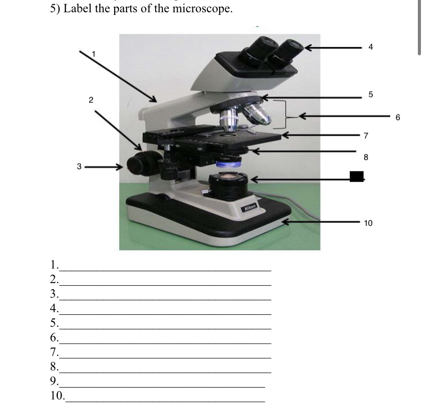
Answered: 5) Label the parts of the microscope. 1… | bartleby

Compound and Stereo- microscopes - Microscopes 4 Schools

Dissecting microscopes vs. Compound microscope

Microscopes. (a) Binocular dissecting microscope. (b ...
Simple Microscope - Parts, Functions, Diagram and Labelling ...

Microscope | Dissecting microscope, Microscope, Microscope parts
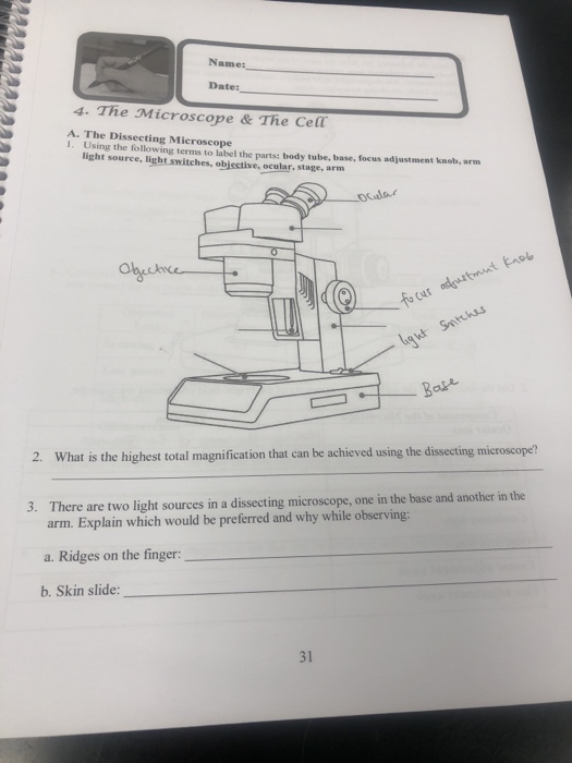
Name: Dates 4. The Microscope & The Cell A. The | Chegg.com

Stereo microscope basics

Simple Microscope - Diagram (Parts labelled), Principle ...

Labelled Microscope with Functions | Microscope parts ...

5 Important Types of Microscopes used in Biology (With Diagram)
Dissection Microscopes
Microscope Review Created by J. Cook. - ppt video online download
Microscopes: Types, parts, magnification, use! - ppt video ...
parts of microscope with diagram - Clip Art Library

Dissecting/Stereo microscope | Principle, Parts, working, and ...

02 Dissecting Microscope. A B Carrying a Microscope. - ppt ...
Label the parts of the microscopes, and also answer | Chegg.com
Lab 1 Introduction

Tsetse biology, systematics and distribution, techniques

Introductory Hydra Activities
Post a Comment for "44 dissecting microscope diagram with labels"